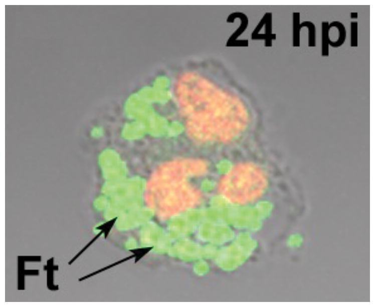
Fig. 2. F. tularensis replicates in PMN cytosol.

The light microcopy image shows a human neutrophil at 24 h post-infection (hpi) with F. tularensis (Ft). DNA in the PMN nucleus was stained red using TOPRO-3. The arrow indicates clusters of green fluorescent protein-expressing bacteria replicating in the cytosol.
