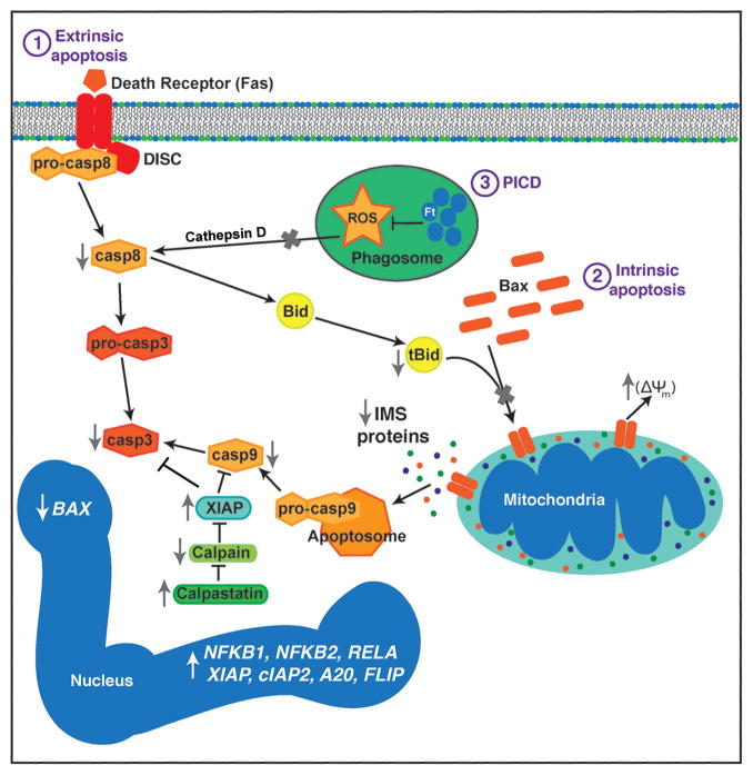
Fig. 3. Mechanisms that contribute to PMN apoptosis inhibition during F. tularensis infection.
The extrinsic, phagocytosis-induced, and intrinsic pathways are shown. F. tularensis inhibits extrinsic pathway activation triggered by Fas crosslinking, whereas blockade of NADPH oxidase activity prevents cathepsin D-mediated caspase-8 activation during PICD. At the same time, Bax translocation to mitochondria and tBID-mediated Bax activation are impaired. Sustained mitochondrial integrity prevents intrinsic pathway activation triggered by intermembrane space (IMS) protein release and apoptosome formation. Calpastatin also contributes to caspase-9 and caspase-3 inhibition by preventing calpain-mediated degradation of XIAP. Effects of F. tularensis on expression of genes that contribute to apoptosis regulation is also depicted. Please see the text for additional details.

