Figure 2.

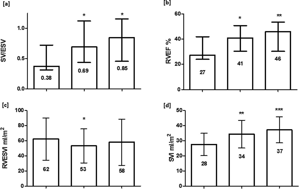
Serial CMR variables for 21 PAH patients performed at diagnosis, 3–8 months and 12–18 months after initiating PAH therapy. median (IQR) or mean (SD) shown. p value in comparison to baseline * p<0.05 ** p<0.01 *** p<0.001 [a]. SV/ESV [b] RVEF [d] SVI increased at 3–8months and were maintained at 12–18 months, one way ANOVA p = 0.006, p = 0.002 and p <0.001 respectively; [c] RVESVI fell at 3–8 months but was unchanged at 12–18 months, ANOVA p = 0.07; no change in RVEDVI occurred (data not shown). RVEF: right ventricular ejection fraction; SV/ESV: RV coupling volumetric method; RVESVI: right ventricular end systolic volume index; RVEDVI: right ventricular end diastolic volume index; SVI: stroke volume index.
