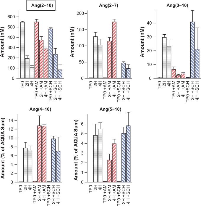
Figure 5.
Normalized angiotensin peak areas from mouse podocytes, incubated with Ang(1-7) indicate production of Ang(2-7) at 1, 2, and 4 hours post addition of substrate (Panel A) and Ang(1-7), incubated with recombinant APA shows production of Ang(2-7) at 60 and 90 minutes that is lost when pretreated with APA inhibitor amastatin (AMA) (Panel B). Ang(1-7) label is inside a text box to indicate that it was the added substrate. Culture media were sampled just following substrate addition (TP0) and 1, 2, 4, and 6 hours post-addition (TP0, 1H, 2H, 4H, 6H). In the absence of cells, the peptide levels were stable over the 6 hour incubation period. Areas were normalized to total angiotensin peptide current and bars show mean of three experiments and one standard deviation about the mean. No evidence of time dependent decay of Ang(1-7), production of other fragments, or post-source decay was evident in the cell-free cases (IC). In-vitro experiments were quantified relative to AQUA Ang(1-7) (500 nmol/L) and AQUA Ang(2-7) (200 nmol/L).

