Figure 2.
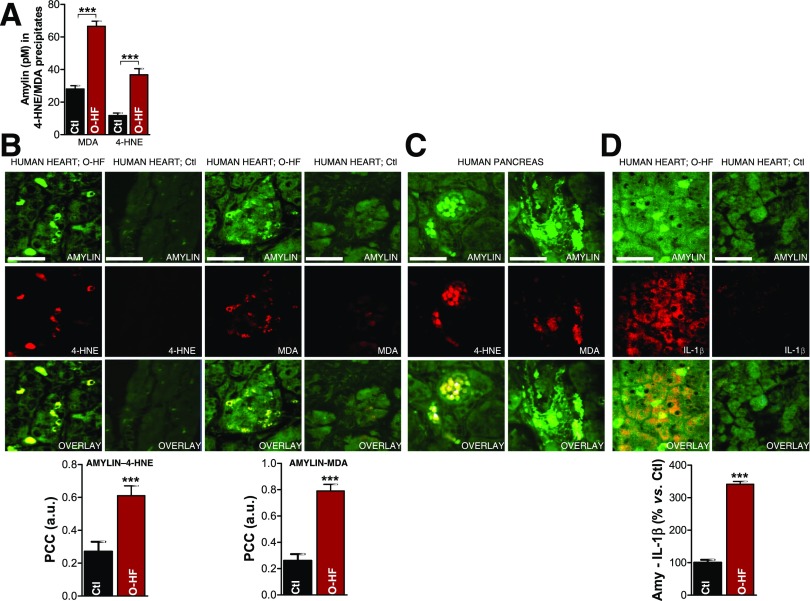
Failing hearts from obese patients display amylin-4-HNE and amylin-MDA adducts and increased IL-1β synthesis. A: 4-HNE and MDA were immunoprecipitated from heart homogenates. The level of amylin in the fractions enriched in 4-HNE and MDA was then quantified by ELISA. Dual-immunofluorescence staining of amylin (green) and 4-HNE or MDA (red) in the heart (B) and pancreas (C) of patients with O-HF (B), patients with type 2 diabetes (C), and Ctl subjects without diabetes (Ctl; B). Bar graphs in B display the mean pixel-by-pixel covariance in amylin and 4-HNE or MDA staining (Pearson’s correlation coefficient [PCC]) in hearts from patients with O-HF vs. Ctl subjects. Scale bars, 20 μm. Ten sections per sample from n = 4 individuals in each group were investigated. D: Immunofluorescence staining of amylin (green) and IL-1β (red) in the heart specimens from O-HF patients and Ctl subjects. Bar graphs show the number of myocytes that are positive for both amylin and IL-1β in a 134 × 134 μm area in heart sections from patients with O-HF vs. Ctl subjects. Scale bars, 20 μm. Ten sections per sample from n = 4 individuals in each group were investigated. Data are presented as the mean ± SE. ***P < 0.001. a.u., arbitrary units.

