Figure 3.
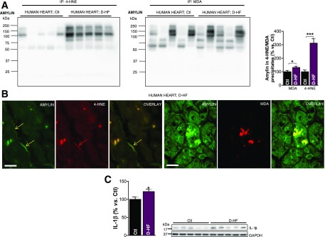
Failing hearts from patients with type 2 diabetes display amylin-4-HNE and amylin-MDA adducts and increased IL-1β synthesis. A: Western blot analysis of enriched 4-HNE and MDA fractions immunoprecipitated from the left ventricles of patients with D-HF vs. nondiabetic Ctl subjects. B: First three images show dual-immunofluorescence staining of amylin (green) and 4-HNE (red) on transverse sections from left ventricle tissue of patients with D-HF vs. nondiabetic Ctl subjects. Arrows indicate amylin incorporation within the sarcolemma and the subsequent formation of adducts with 4-HNE. The next three images are cross sections of cardiac tissue showing the formation of amylin-MDA adducts within cardiac myocytes. Scale bars, 20 μm. C: Western blot analysis of IL-1β in left ventricle tissue of patients with D-HF vs. nondiabetic Ctl subjects. A representative blot from two independent experiments is shown. Data are presented as the mean ± SE. *P < 0.05; ***P < 0.001.

