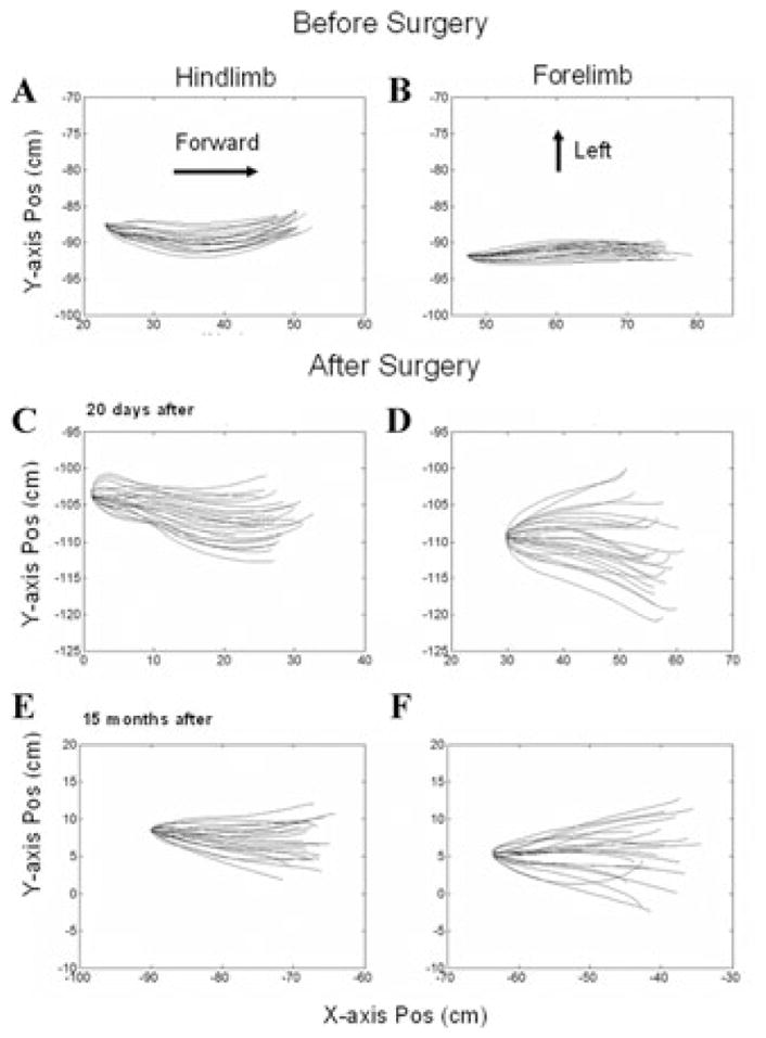
Figure 3.

Swing-phase trajectories of right hindlimb (A, C, E) and right forelimb (B, D, F) before (A, B), 20 days after (C, D), and 15 months after (E, F) all six semicircular canals were plugged. The swing phases were normalized to the same initial position, and the trajectories are displayed in the X–Y plane (top view). The displacement in the X direction is shown on the abscissae, and the Y direction on the ordinates. Note the marked splaying of the trajectories after surgery.
