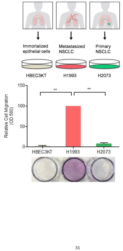
Figure 1. H1993 cells are associated with a metastatic phenotype.
HBEC3-KT, H1993 and H2073 cells were cultured in serum-free media in the insert of a transwell apparatus. The lower chamber was filled with media containing FBS. After 48 hours, cells migrated through the membrane were stained with crystal violet. The crystal violet was then dissolved with 33% acetic acid and the absorbance at 560 nm was measured from three independent experiments (**, p < 0.01).

