Figure 6.
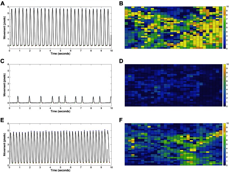
Knockdown of DNMT3a expression increases beating rate variability and decreases movement in embryonic cardiomyocytes. A, C, E) The trace of cell movement from the video-based imaging assay revealed that transient knockdown of DNMT3a expression for 72 h in cardiomyocytes decreased beating frequency and cell movement and increased beating rate variability (C) compared with the cells treated with negative siRNA (A) or DNMT3b siRNA (E). B, D, F) The heatmap of cell movement across the region of interest also indicated less contractile movement in the DNMT3a siRNA–treated cardiomyocytes (D) than that in negative siRNA (B) or DNMT3b siRNA–treated cells (F). Yellow, high-movement pixels; blue, low-movement pixels.

