Figure 1.
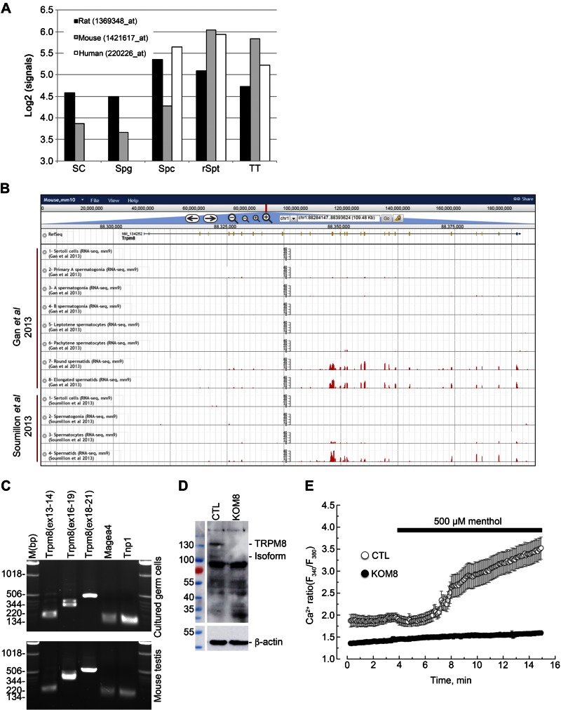
TRPM8 channels are expressed and functional in mouse germ cells. A) Normalized signal intensities (y axis, log2-transformed) of Trpm8 are shown in the different testicular cell types (x axis) in Sertoli cells (SC), spermatogonia (Spg), spermatocytes (Spc), round spermatids (rSpt), and total testis (TT) of 3 mammalian species: rat, mouse, and human. B) Gene structure is shown for Trpm8 and histograms of the numbers of RNA-seq reads that aligned the corresponding genomic locations across the different samples from Gan et al. (21) and Soumillon et al. (22) (y-axis ranges from 0 to 40) (adapted from The ReproGenomics Viewer; http://rgv.genouest.org/publication.html). C) PCR detection of different regions of Trpm8 in cultured germ cells (top) and in whole extracts of mouse testis (bottom). PCR fragments were amplified from exon X to Y and reported as Trpm8(exX-Y). Melanoma antigen family A-4 (Magea 4) and transition protein 1 (Tnp1) were used as reporters of spermatogonia and spermatids respectively. D) Immunoblot analysis reveals detection of full-length TRPM8 (130 kDa) and a 105 kDa TRPM8 isoform, as well, in total protein extract of CTL mouse testis, but not in TRPM8-KO (KOM8) mouse testis. E) Calcium imaging experiments realized with Fura2-AM fluorescent probe show an increased cytosolic Ca2+ concentration in 2 d isolated Trpm8+/+ mouse germ cells (CTL; n = 20) after addition of 500 µM menthol. No Ca2+ variation was detected in germ cells of Trpm8−/− mouse line (KOM8; n = 83).

