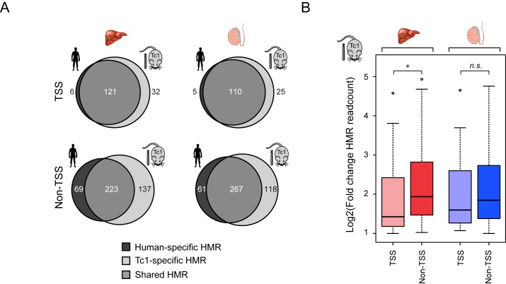
Figure 3.
The majority of species-specific HMRs are distal to gene promoter regions. (A) Venn diagrams depicting the overlap between HMRs in the human and Tc1 mouse tissue. HMRs are segregated into those located at transcription start site (TSS ± 500bp) (upper) and those found away from TSSs (lower). Human-specific HMRs are coloured in dark grey, shared HMRs in grey and Tc1-specific HMRs in light grey. (B) Boxplots depicting fold-change in BioCAP signal between Tc1 mouse and human at Tc1-specific HMRs located at a gene TSS or elsewhere in the chromosome. Tc1-specific HMRs away from gene TSSs exhibit a greater fold-change in BioCAP signal compared to those at gene TSSs. Significance values calculated using Welch's T-test (*P < 0.05).

