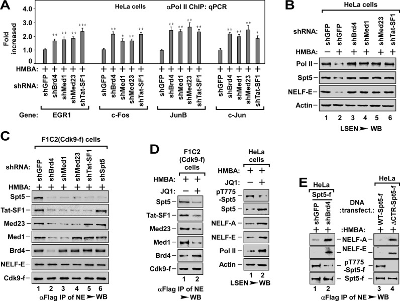
Figure 3.
The P-TEFb targeting DSIF is recruited by Brd4 via a recruitment pathway consisting of Med1, Med23 and Tat-SF1. (A) The Pol II-bound genomic DNAs ChIPed from HeLa cells with indicated shRNA infection and HMBA treatment were analyzed by qPCR for the enrichment of Pol II on the promoter region of representative genes as in Figure 1E. The level in shGFP infected cells was set to 1.0. All values were expressed as Mean ± SD of three replicates. *P < 0.05, **P < 0.01, ***P < 0.001; P-values were assessed using two-tailed Student's t-test. (B) LSEN from HeLa cells with indicated shRNA infection and HMBA treatment were analyzed by WB as in Figure 1C. (C) Effect of depletion of Brd4, Med1, Med23 or Tat-SF1 on the recruitment of P-TEFb to DSIF (Spt5) and NELF (-E). Anti-Flag immunoprecipitates from NEs of F1C2 (Cdk9-f) cells with indicated shRNA infection and HMBA treatment were analyzed by WB for the indicated Cdk9-f/P-TEFb-associated proteins. (D) Effect of JQ1 on P-TEFb recruitment to Spt5/DSIF (left panel) and P-TEFb-mediated phosphorylation of Spt5 (pT775-Spt5, right panel). The anti-Flag immunoprecipitates from NEs of F1C2 (Cdk9-f) (left panel) and the cell lysates of HeLa cells (right panel) with indicated JQ1 and HMBA treatment were analyzed by WB for the indicated proteins. (E) Effect of Spt5 phosphorylation on the dissociation of NELF. The anti-Flag immunoprecipitates from NEs of HeLa cells with indicated Spt5-f cDNA and shRNA co-transfection (left) or with WT- or ΔCTR-Spt5-f transfection (right) were analyzed by WB for the levels of NELF-A and NELF-E and the levels of phosphorylated Spt5 (pT775-Spt5).

