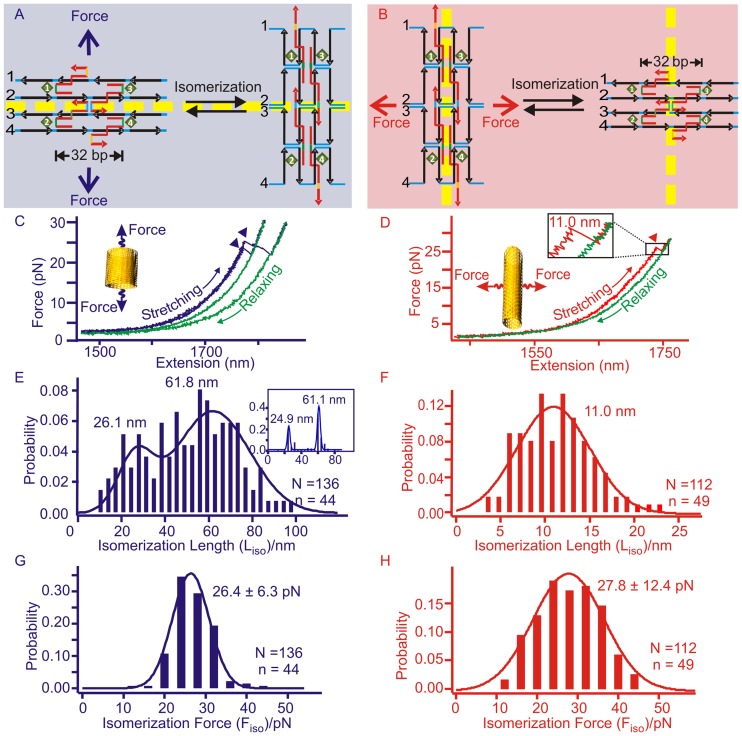
Figure 4.
Mechanical isomerization of the eight-tube DNA origami. Mechanical isomerizations between the long and short DNA tubes are explained from the perspective of Holliday junctions in the origami structure during longitudinal (A) or horizontal (B) stretching. For clarity, only one complete Holliday junction is shown in the middle of each structure. Dotted yellow lines depict Holliday junction layers in different stretching orientations. Representative F-X curves were obtained during longitudinal (C) or horizontal (D) stretching of the eight-tube DNA. Arrow heads in both F-X curves indicate the isomerization events (see insets for respective schematic diagrams). The blue/red and green traces depict stretching and relaxing curves, respectively. Due to the nature of the mechanical isomerization, only the transitions from the short tube to the long tube in the longitudinal pulling, and the long tube to the short tube in the horizontal pulling can be observed. Histograms of the isomerization length (Liso) of the eight-tube DNA are plotted for longitudinal (E) or horizontal (F) stretching (inset in E depicts the most probable populations using PoDNano approach, see text). Histograms of the isomerization force (Fiso) of the same DNA tube measured during longitudinal (G) or horizontal (H) stretching. Solid curves represent Gaussian fittings. N and n respectively depict the number of F–X traces and number of molecules in experiments.

