
Fig 4.
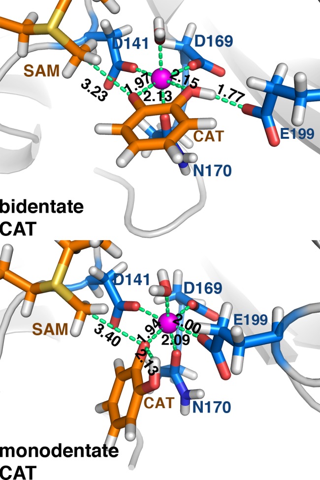
Representative bidentate (top) and monodentate (bottom) catecholate (CAT) substrate configurations at the COMT active site. Substrates are shown in orange, protein residues in blue, and key distances are shown (in Å), except for D141-Mg2+, D169-Mg2+, and Mg2+-H2O, which are unchanged.