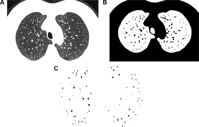
Figure 1.
Measurement of cross-sectional area of small pulmonary vessels using ImageJ software in normal subjects.
Notes: (A) CT image of lung field segmented within the threshold values from −500 to −1,024 HU. (B) Binary image converted with window level of −720 HU from segmented image. (C) Pulmonary vessels are displayed in black.
Abbreviation: CT, computed tomography.

