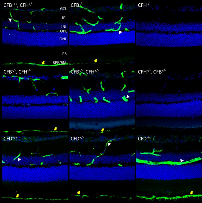
Fig 3. Immunostaining of C3 is restored to wild type levels in the retinal vessels and RPE/Bruch’s space in Cfh-/- mice when in the genetic background of Cfb-/- but not Cfb+/-.

12 μm PFA fixed sections were taken of mouse retinas of the indicated genotypes at age 12 months. Sections were stained for C3-FITC (green) and nuclei (blue). Yellow arrows highlight staining in RPE/Bruch’s. Z-stacks were imaged using confocal microscopy and merged to form a maximum intensity projection. Scale bar = 50 μm. Images are representative of n = 6.
