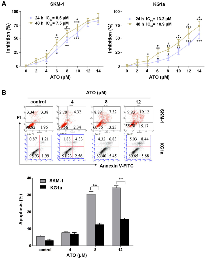
Figure 2.
ATO inhibits cell growth and induces cell apoptosis in SKM-1 and KG1a cells. (A) SKM-1 and KG1a cells were exposed to different concentrations of ATO for 24 and 48 h. The inhibitory rates of the cells were detected by MTT assays. The IC50 value of ATO in the two cell lines are shown. (B) SKM-1 and KG1a cells were treated with different concentrations of ATO for 48 h, and then apoptotic cells were analyzed by staining with Annexin V/PI. The MTT and apoptosis results showed a dose-dependent effect in the two cell lines. The data are expressed as the means ± SD of three independent experiments; *p<0.05, **p<0.01, ***p<0.001 (compared with the control); #p<0.05 (24 vs. 48 h).

