Figure 1.
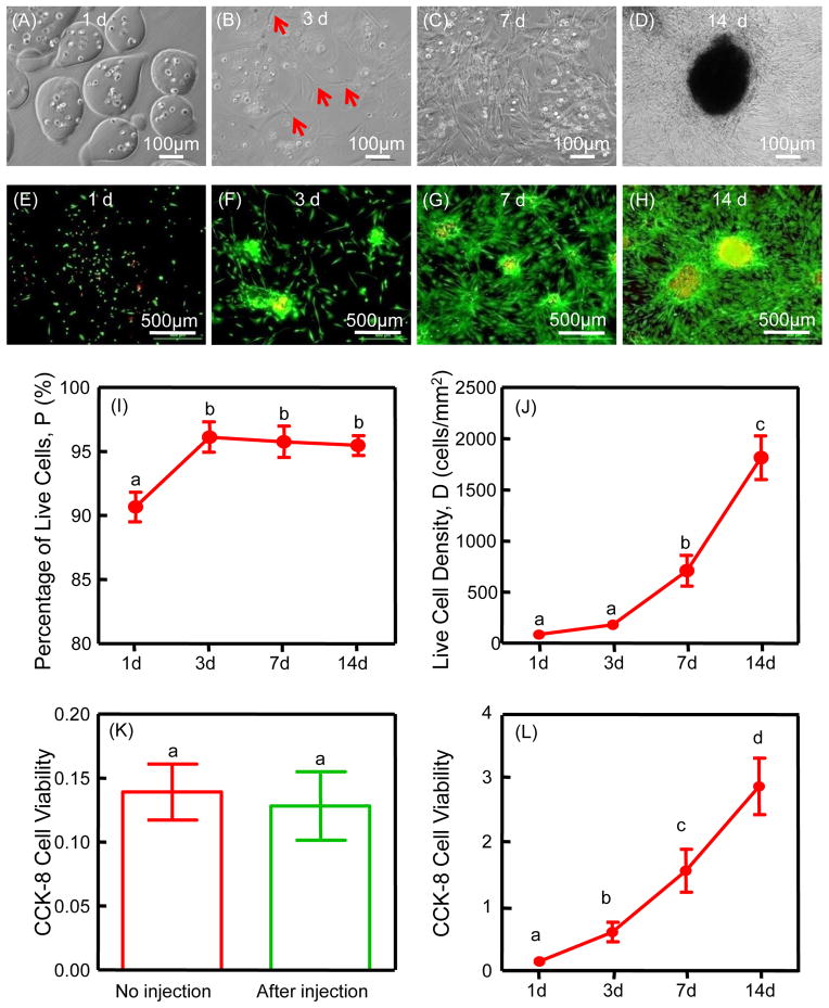
Cell encapsulation and release from the microbeads (A–D): at 3 d, iPSMSCs started to be released from microbeads; from 7 to 14 d, more cells were released and the contour of microbeads became obscure as alginate degraded. Cell viability and proliferation of iPSMSC-microbeads (E–L): live/dead staining and CCK-8 from 1 to 14 d showed good cell viability and proliferation, and cell viability (absorbance at 450 nm) was not negatively affected by injection (K). CCK-8 cell viability (absorbance at 450 nm) increased with culture time (L). Each value is mean ± SD (n = 6). Bars with dissimilar letters indicate signi cantly different values (p < 0.05).

