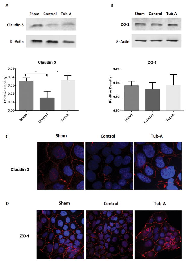
Figure 5.
Tubastatin-A protects claudin-3 protein from loss in Caco-2 cells in anoxia condition. Representative western blots and densitometry analysis (A, B) as well as immunofluorescence staining (C, D) of claudin-3 and ZO-1 in Caco-2 cells treated with different conditions. Control cells showed significant decrease of claudin-3 compared with sham (*P=0.003), Tubastatin-A treatment protected claudin-3 from loss (*P=0.002 vs. control, A). No significant difference in the expression of ZO-1 protein between the sham, control, and Tubastatin-A treatment groups (B). Normal Caco-2 cells had strong membrane and weak cytoplasmic signals of claudin-3 protein, anoxia caused pronounced loss, while treatment with Tubastatin-A increased the stainings (C). ZO-1 protein strongly expressed in the cellular membrane, while anoxia didn’t result in significant loss and Tubastatin-A treated cells exhibited a moderate change compared to control cells (D). Values are expressed as mean ± SD from three independent experiments. Original magnification 200×; red color: claudin-3 and ZO-1 signal; blue color: nuclear Hoechst staining. Sham: no anoxia injury; Control: anoxia without treatment; Tub-A: Tubastatin-A treatment.

