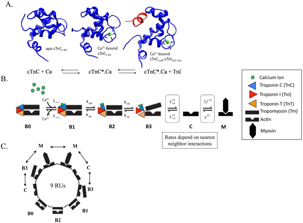
Figure 2.
(A) Molecular state of cTnC (in blue) in apo state (BO in Markov model), calcium (in green) bound state (B1 in Markov model) and calcium-bound state with exposed hydrophobic patch allowing for cTnI (in red) to bind to cTnC (B2 in Markov model). (B) Schematic of one regulatory unit (RU) sequence of the six-state (BO, B1, B2, B3, C, M) Markov model of cooperative myofilament activation with nearest neighbor interactions. (C) Schematic of ring structure mathematical formulation used to solve the Markov model that incorporates looping of short segments of RUs to allow elimination of redundant states due to symmetry, resulting from the tight co-operative coupling of 9 adjacent RUs, linked by tropomyosins, depicted in one possible configuration of nearest neighbor interactions (the double-headed arrow) (see Methods and26 for more details). The nearest neighbor interactions of one RU with two adjacent RUs, X and Y – which could be in either Markov state B3, C or M (as in 26), determine the degree of co-operativity of myofilament activation in the Markov model.

