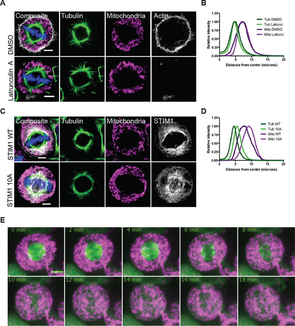
Figure 2. Mitochondrial distribution is independent of actin or ER tethering during mitosis.
(A) HeLa cells were synchronized into metaphase cells and treated for 10 min with DMSO or Latrunculin A prior to fixation. Mitochondria (TOM20, magenta), tubulin (green), and actin filaments (grey) were immunostained and imaged by confocal microscopy. (B) The radial distributions of tubulin and mitochondria were averaged for 30 metaphase cells treated as in (A). (C) HeLa cells were transiently transfected with STIM1 constructs. Mitochondria (TOM20, magenta), tubulin (green), and STIM1 (grey) were immunostained and imaged by confocal microscopy. (D) The radial distributions of tubulin and mitochondria were averaged for 30 metaphase cells expressing STIM1 WT or STIM 10A as in (C). (E) HeLa cells were transfected with Mito-dsRed and GFP-tubulin and synchronized. Cells were treated with nocodazole (0 min) and images were taken every 2 minutes. Scale bars represent 5 microns.

