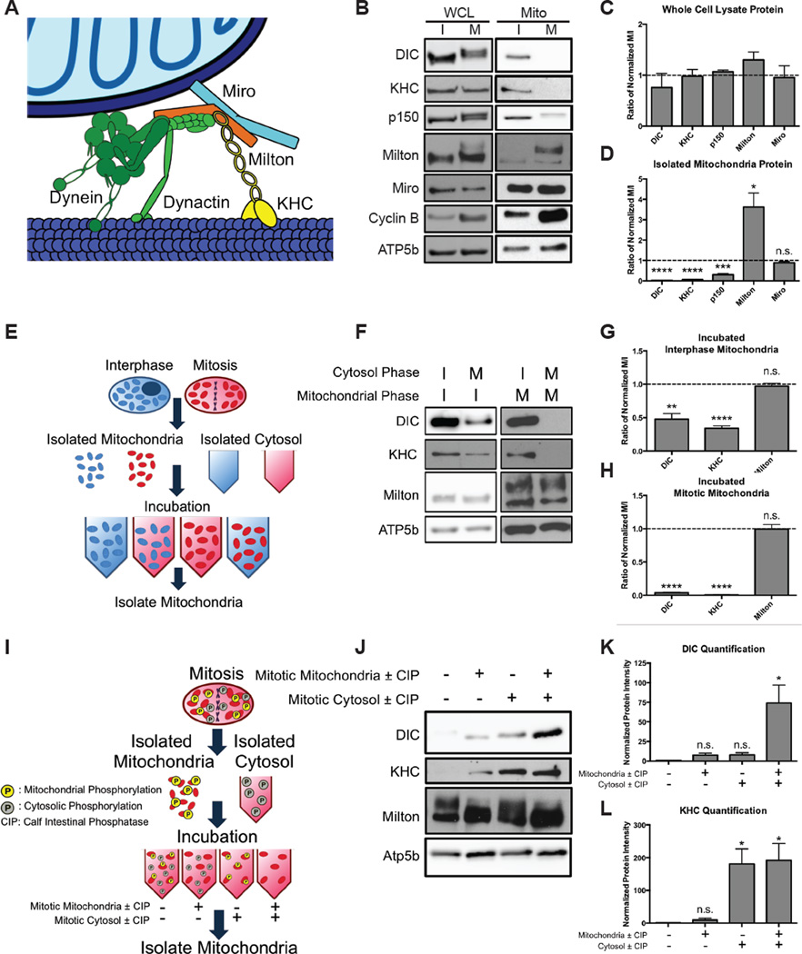
Figure 3. Phosphorylation detaches dynein and kinesin motors from mitochondria during mitosis.
(A) Schematic of the motor adaptor complex including Miro, Milton, kinesin (KHC), dynein and dynactin. (B–D) HeLa cells were synchronized into interphase or mitosis (nocodazole-induced arrest). Whole cell lysates (WCL) and isolated mitochondria (Mito) were probed for the indicated proteins of the motor adaptor complex. Levels of the motor protein subunits were reduced on mitotic mitochondria, but Milton and Miro remained on mitochondria. Several protein’s positions were altered by mitotic phosphorylations. Elevated Cyclin B levels verified that cells were in mitosis, and the mitochondrial protein ATP5b verified equal mitochondrial content in the samples. Band intensities were quantified (C,D), and each mitotic protein was normalized to the level of that protein at interphase and the fold changes are shown. n = 3. (E–H) Schematic, immunoblot, and quantification of a biochemical assay to determine if interphase or mitotic cytosol can alter the mitochondrial association of the motors. Mitochondria from either interphase (I) or mitosis (M) were incubated with interphase or mitotic cytosol. Mitochondria were re-isolated and assayed for the indicated proteins. Mitotic cytosol induced motor release, and interphase cytosol reattached motors in the assay. Band intensities were quantified, and the relative effects of the two types of cytosol were compared by normalizing the intensity of the band with mitotic cytosol to that with interphase cytosol. The fold changes for interphase mitochondria (G) and mitotic mitochondria (H) so treated are shown. n = 3. (I–L) Assay to determine if treatment with calf intestinal phosphatase (CIP) can allow motors to reattach. The mitochondrial and cytosolic fractions were treated with either CIP alone (+) or CIP and the phosphatase inhibitor NaVO4 (−) prior to being recombined. Mitochondria were re-isolated and probed for the indicated proteins. DIC (K) and KHC (L) levels were quantified and normalized to the untreated fraction condition. n = 3. *p < 0.05, **p < 0.01, ***p < 0.001, ****p < 0.0001.

