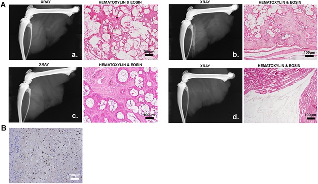
Figure 2.

Human mesenchymal stem cells (hMSCs) were unable to rescue HO. (A) Radiographic and histological analysis of tissues isolated 2 weeks after induction of HO in the presence and absence of hMSCs. Wistar rats were injected at the distal (a) or proximal (b) location with AdBMP2 transduced cells and the distal location with hMSCs + AdBMP2 transduced cells (c) or hMSCs + PBS (d). (B) Photomicrograph of mouse tissue isolated 2 weeks after induction of HO by delivery of AdBMP2 transduced cells in the presence of hMSCs. The differentiation of the hMSCs into mesenchymal tissues of bone was confirmed through immunostaining with an antibody that specifically detects the human cells (brown). The representative section shows the presence of human chondrocytes within the mouse bone.
