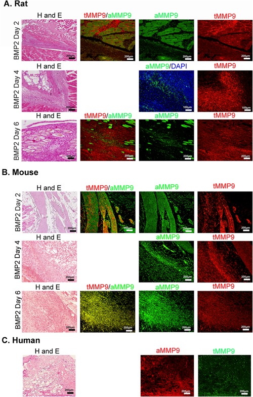
Figure 4.

Expression of MMP9 active and inactive forms within the area of HO. Representative photomicrographs of paraffin sections serially cut from rat (A), mouse (B), or human (C) tissues. Rat and mouse tissues were isolated 2, 4, and 6 days after delivery of AdBMP2 transduced cells. Human tissues depict soft tissues immediately surrounding the newly forming heterotopic bone. Tissue sections were stained with hematoxylin and eosin or co‐immunostained with antibodies that detect total MMP9 (red) or the active form (green), or for human tissues total MMP9 (green) and active form (red) as indicated.
