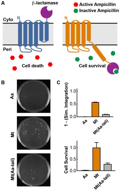
Figure 4. Correlation of antibiotic resistance to membrane topology.
(A) Schematic of the cytoplasmic and periplasmic topologies of the TatC C-tail with the fused β-lactamase enzyme. Misintegration of loop 7 leads to periplasmic localization of the β-lactamase, resulting in enhanced antibiotic resistance and cell survival. (B) Representative plates from the ampicillin survival test. (C) Comparison of the simulated integration efficiency (top) and relative ampicillin survival rate (bottom) for AaTatC, MtTatC, and Mt(Aa-tail). The reported cell survival corresponds to the ratio of counted cells post-treatment versus prior to treatment with ampicillin; all values are reported relative to MtTatC. Error bars indicate the standard error of mean.

