Fig. 1.
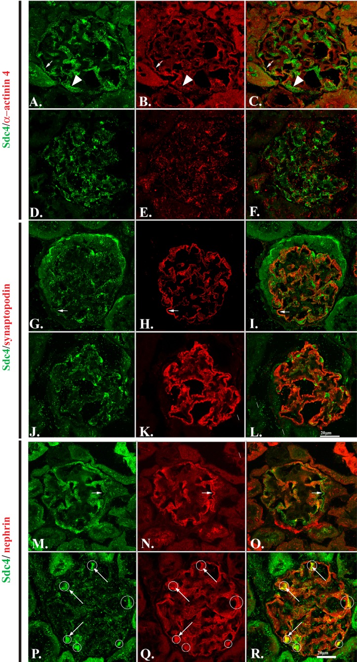
Podocytes from Ndst1−/− mice show disruption in syndecan-4 (Sdc-4) distribution and alterations in cytoskeletal organization in vivo. A–C, G–I, and M–O: laser scanning confocal micrographs of kidneys from Ndst1+/+ animals at 15 mo of age. D–F, J–L, and P–R: micrographs of kidneys from Ndst1−/− mice of the same ages. This age range was selected because microalbuminuria is evident at this age in the Ndst1−/− mouse model [see (42)]. The sections were stained for Sdc-4 (A, D, G, J, M, and P) and α-actinin-4 (B and E), synaptopodin (H and K), and nephrin (N and Q). C, F, I, L, O, and R: overlay images of the panels within the same row of images. The data show that the glomerular podocytes in Ndst1−/− mice show significant changes in the organization/localization of Sdc-4 (D, J, and P). Of the three molecules commonly associated with glomerular podocytes, the localization of α-actinin-4 (E) is most disrupted in Ndst1−/− podocytes compared with that of controls (B). The pattern of synaptopodin staining is also changed (H and K), albeit not to the same extent observed with α-actinin-4 (B and E). The pattern of nephrin staining (N and Q) appeared relatively the same in the glomeruli from both strains of mice, with the only difference being the colocalization/coclustering of Sdc-4/nephrin in aggregates (R, arrows) in the glomeruli of Ndst1−/− mice. Neither synaptopodin nor α-actinin-4 show a similar pattern of colocalization in the glomeruli of Ndst1−/− mice (final magnification ×400, bar = 20 μm).

