Fig. 2.
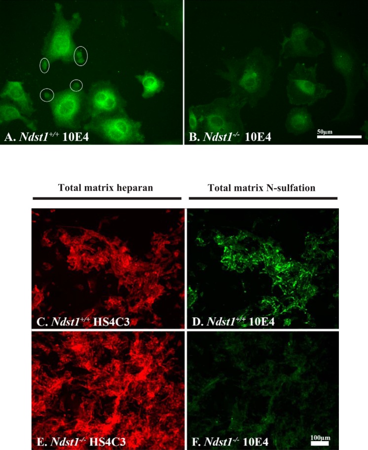
Ndst1−/− podocytes are unable to N-sulfate heparan sulfate (HS) glycosaminoglycans. A and B: Ndst1+/+ and Ndst1−/− podocytes grown in short-term cell culture, and the cells are immunostained with the 10E4 antibody to demonstrate N-sulfated residues. On the periphery of the Ndst1+/+ cells, “pods” of 10E4-positive immunoreactivity (white circles) can be seen around the periphery of the cells, whereas in Ndst1−/− cells (B) there is little to no 10E4 staining at the cell periphery. The perinuclear staining observed in Ndst1−/− cells for 10E4 is the result of nonspecific staining of the secondary antibody. For comparative purposes, both images were shot at the same exposure and subjected to the same normalization with regard to gray-level intensity. C–F: Ndst1+/+ and Ndst1−/− podocytes grown in culture for 5 days, and cultures were subsequently immunostained for the presence of heparin sulfate (HS) (C: E-HS4C3 antibody) or the presence of N-sulfated residues (D: F-10E4 antibody). For comparative purposes, the exposures in C and E were identical as were exposures in D and F. Each image set was subjected to the same normalization with regard to gray-level intensity. C and D: Ndst1+/+ podocytes are able to assemble a pericellular matrix containing N-sulfated (D) HS (C); the Ndst1−/− podocytes are able to assemble a pericullar matrix containing HS (E) but with little or no N-sulfation (F). Final magnification: A and B, ×400, bar = 50 μm; C–F, ×100, bar = 100 μm.

