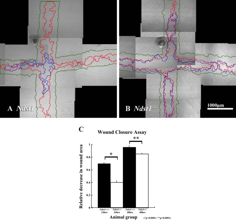
Fig. 5.
Ndst1−/− podocytes migrate less efficiently compared with Ndst1+/+ podocytes. The micrographs show representative fields of view of wounded monolayers of Ndst1+/+ (A) and Ndst1−/− (B) podocytes. In each micrograph, the monolayer boundaries at a given postwounding time interval are demarcated by green lines (T = 0), red lines (T = 24 h), and blue lines (T = 48 h). The white asterisk in each micrograph denotes one of several marks that were used as registration points for image alignment. C: differences in respective areas (A) for 24 h (A0-A24) and 48 h (A0-A48). The data show that at each time interval the amount of wound closure is significantly greater (P = 0.001) in the Ndst1+/+ cells compared with the Ndst1−/− cells. Final magnification in A and B = ×40, bar = 1,000 μm.

