Fig. 6.
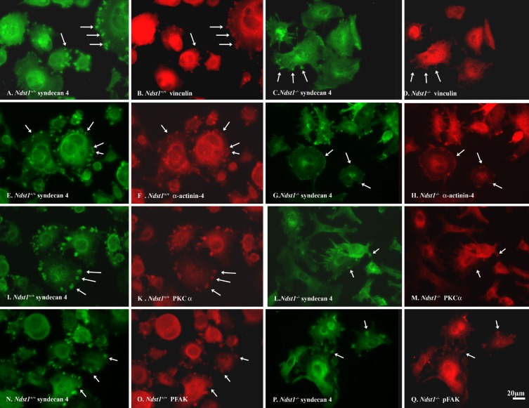
Ndst1−/− podocytes do not readily form adhesion complexes on fibronectin compared with Ndst1+/+ podocytes. A, B, E, F, I, K, N, and O: micrographs of Ndst1+/+ podocytes; C, D, G, H, L, M, P, and Q: micrographs of Ndst1−/− podocytes immunostained for Sdc-4 (A, C, E, G, I, L, N, and P), vinculin (B and D), α-actinin-4 (F and H), PKCα (K and M), or pFAK (O and Q). In Ndst1+/+ cells, Sdc-4 staining appears as large aggregates (“pods”) at the periphery of the cells (arrows in A, E, I, and N). In Ndst1−/− cells (C, G, L, and P) the aggregates are smaller in size. In accord, in Ndst1+/+ cells, the relative size of vinculin (B), α-actinin-4 (F), PKCα (K), and pFAK (O) clusters are larger than those observed in Ndst1−/− cells (D, H, M, and Q, respectively). Final magnification ×400, bar = 20 μm.

