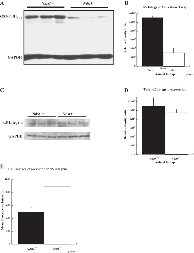
Fig. 8.
The presence of α5β1 integrin in the active state at the cell surface in Ndst1−/− podocytes is diminished compared with that observed on the surface of Ndst1+/+ podocytes. A: Western immunoblot for GST-tagged FNIII9–11 peptide that had been bound to the activated α5β1 integrin on the surfaces of Ndst1+/+ and Ndst1−/− podocytes. B: graph of the densitometry of the respective bands of the gel shown in (A), the measures normalized to the GAPDH loading control. The amount of GST-FNIII9–11 peptide bound to activated integrin is significantly less (P = 0.023) in the Ndst1−/− cells compared with the Ndst1+/+ cells. C: Western blot of total cellular α5 integrin expression of Ndst1+/+ and Ndst1−/− podocytes. D: densitometry measures of the blot in (C) normalized to the GAPDH loading control. The data show that there was not a significant difference in the levels of total α5β1 integrin expression between of Ndst1+/+ and Ndst1−/− podocytes. E: results of surface immunostaining/flow cytometry measurements of Ndst1+/+ and Ndst1−/− podocytes for the presence of α5 integrin subunit at the cell surface. The data show Ndst1−/− podocytes that have more α5 integrin on their surfaces than Ndst1+/+ cells. Taken together, the data indicate that Ndst1−/− podocytes have more α5 integrin on their surfaces but is held in the low-affinity state for ligand binding.

