Fig. 2.

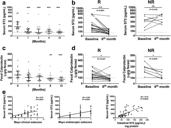
Distribution of serum ST2 according to response to therapy. a Serum ST2 levels during the follow-up period were determined in each patient and are shown as one symbol in the scatter plot. Horizontal lines indicate medians and whiskers (interquartile ranges). Differences were assessed using Kruskal-Wallis test (*** p < 0.0001). b Serum ST2 levels at baseline and 6 months in responders (R) and non-responders (NR) in relation to therapy decreased only in responders. Differences were assessed using Wilcoxon signed rank test (*** p < 0.0001). c FC levels in the 1-year follow-up and (d) at baseline and 6 months in R and NR in relation to therapy decreased in both subgroups. Panel E shows the correlation between serum ST2 levels and Mayo clinical subscore, Mayo endoscopic subscore, and total ST2 intestinal mucosa content, with a trend line for each correlation. Discontinuous line indicates cut-off value. Rs: Spearman’s rank correlation coefficient
