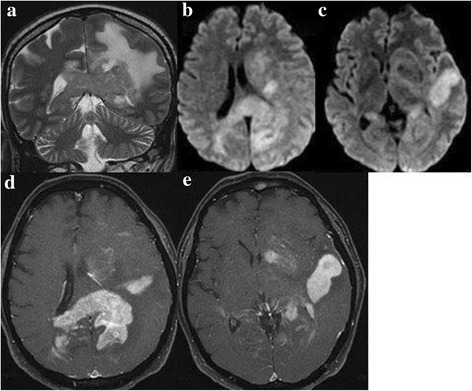
Fig. 1.

MRI findings strongly suggestive for brain lymphoma: SE T2 sequences in coronal plane (a), diffusion in axial plane (b, c) and fat-suppressed T1 after contrast medium infusion in axial plane (d, e). MRI shows multiple bilateral, periventricula sites with low signal intensity on T2, with oedema, hyperintensity signal on DWI and homogensously enhancement after contrast medium infusion
