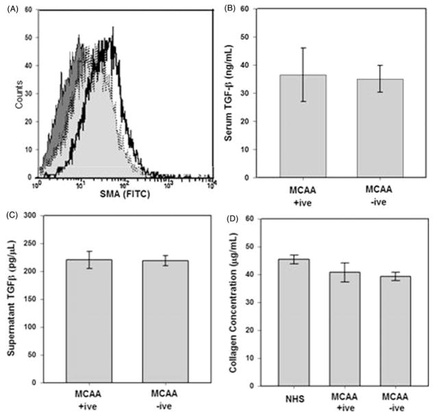
Figure 3.
MCAA exposure does not induce a mesenchymal transition in cultured human pleural mesothelial cells. (A) SMA expression was measured in cells exposed to human sera or TGF-β (5 ng) as a positive control for 4 days. Cells were fixed and stained with primary anti-SMA and secondary FITC conjugated antibodies and mean fluorescence measured by flow cytometry. TGF-β (
 ) induced SMA expression compared with exposure to secondary antibody control (
 ,
,
 ), MCAA +ive (
 ,
,
 ), or MCAA −ive (
 ) sera. Exposure to MCAA +ive or −ive sera resulted in increased SMA expression over secondary control (x-axis) but expression did not increase to the same degree as TGF-β exposure. TGF-β was detected in MCAA +ive and −ive sera (B) and supernatants (C) of cells exposed to these sera samples. TGF-β was detected using a cytokine bead array analysis and concentrations suggest that the TGF-β detected in supernatants was added upon addition of sera and not produced by the mesothelial cells, mean ± SD, n =3). (D) Total soluble collagen concentrations in cell supernatants were determined using a Sircol Soluble Collagen Assay. Incubation with sera from asbestos-exposed subjects did not significantly affect soluble collagen compared to NHS.

