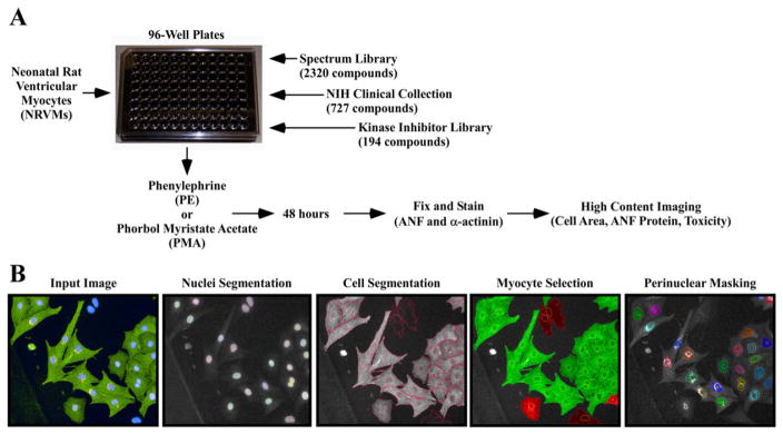
Fig. 1.
A novel high throughput assay of cardiomyocyte hypertrophy. (A) Neonatal rat ventricular cardiomyocytes (NRVMs) were seeded on 96-well plates and treated with candidate compounds in the presence of PE or PMA for 48 h. Cells were fixed, subjected to indirect immunofluorescence with antibodies against ANF and α-actinin, and stained with Hoechst dye prior to high content imaging. (B) Images were analyzed using Harmony software. Cells were identified based on nuclei segmentation (Hoechst), and subsequently individual cells were segmented and cell area was calculated based on area of α-actinin fluorescence (FITC). Non-myocytes were removed from analysis based on the absence of α-actinin signal. Finally, ANF expression was measured using a perinuclear mask (Cy3).

