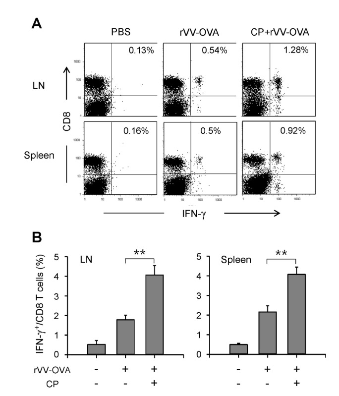
Figure 5. CP increased IFN-γ producing CD8 T cells in mice infected with rVV-OVA. Mice were orally administered CP, infected with VV-OVA, and then treated with the same doses of CP for consecutive 5 days, as described in Fig. 1. On the final day of CP administration, CFSE-labeled OT-I cells were injected through the tail vein. After 4 days, the lymph node and spleen cells were isolated and restimulated for 6 h with OVA[257-264] peptide-pulsed DCs. The cells were stained with an anti-CD8 mAb, then permeabilized and stained with an anti-IFN-γ mAb. (A) Representative histograms are shown. (B) The percentages of IFN-γ+ cells among total CD8+ T cells are shown graphically (n=5 for each group). **p<0.01.

