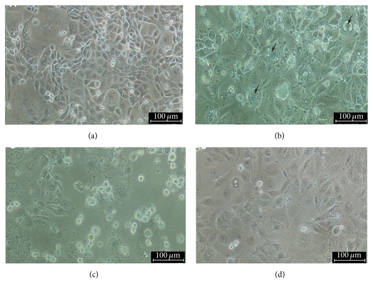
Figure 2.
Infection of MDCK cells with virus suspension revealed evidences of virus growth as demonstrated by CPE which is characterized by cell ballooning, granulation, formation of giant cells (arrow), rounding, and cell detachment as infection progresses 3 dpi (a); 5 dpi (b) and 7 dpi (c); (d) control uninfected cells. Mag ×20 and 100 μm.

