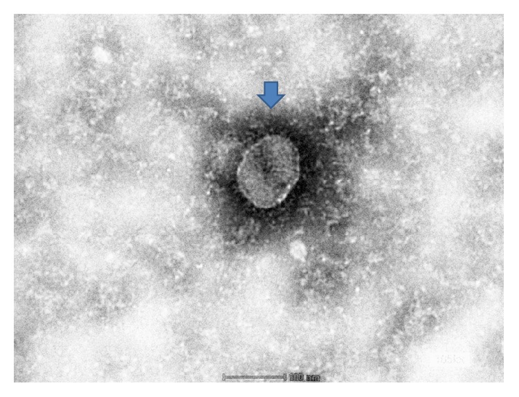
Figure 3.

Negative staining electron microscopy showing the morphological appearance of the isolated virus. Note: presence of spike projections surrounding the viral particle (arrow) whose diameter ranges from 80 to 120 nm. Mag ×100 μm.

Negative staining electron microscopy showing the morphological appearance of the isolated virus. Note: presence of spike projections surrounding the viral particle (arrow) whose diameter ranges from 80 to 120 nm. Mag ×100 μm.