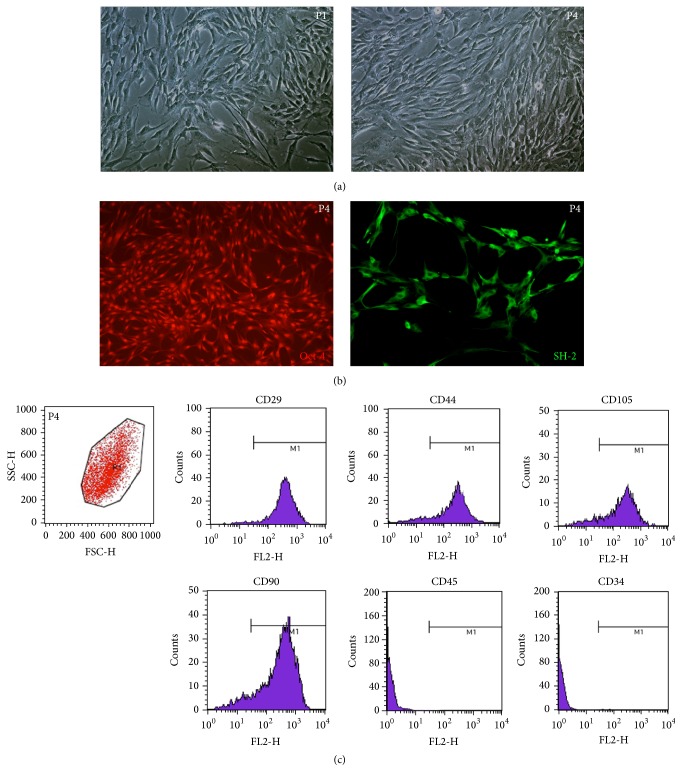
Figure 1.
Morphology and molecular marker detection of isolated ADMSCs. (a) Phase-contrast micrographs of ADMSCs at P1 and P4. Most ADMSCs were spindle-shaped with scant cytoplasm and with granules around the nuclei (original magnification 100x). (b) Immunofluorescence detection: P4 ADMSCs were positive for Oct-4 and SH-2 (original magnification 100x). (c) FACS analysis: P4 ADMSCs were positive for CD29, CD44, CD105, and CD90 and negative for CD45 and CD34.

