
Figure 5.
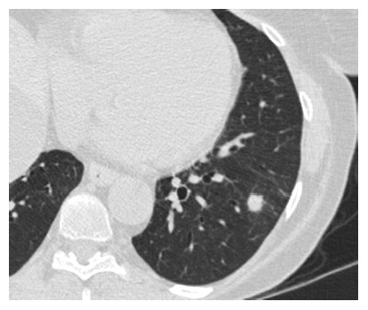
Typical appearance of an indeterminate solid pulmonary nodule at high-resolution computed tomography (Philips icomputed tomography, slice thickness 1.25 mm) in a 59-year-old female patient. The lesion was located in the left lower lobe, had a maximum diameter of 10 mm and proved to be a non-tubercular granuloma at surgery.