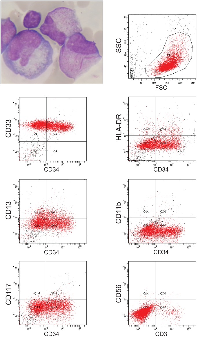
Figure 1.
Clinical manifestations of meningeal APL relapse with Giemsa staining (upper left panel) and fluorescence-activated cell sorting analysis of CD33, CD34, HLA-DR, CD13, CD11b, CD117, CD56, and CD3 expression from cerebrospinal fluid blasts at relapse. Cells were gated in the side scatter (SSC)/forward scatter (FSC) analysis as shown in the upper right panel.

