
Figure 1.
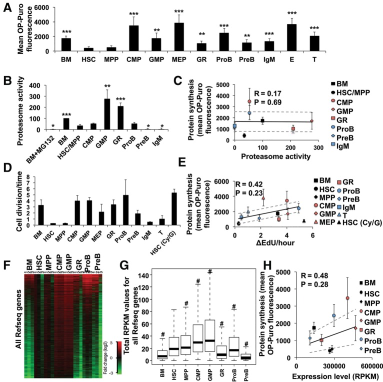
Protein synthesis rates in HSCs and progenitor cells show little correlation with proteasome activity, frequency of cell division, or RNA content. (A) OP-Puro incorporation by HSCs and progenitor cells in vivo. n = 8 mice in eight experiments. (B) Proteasome activity in HSCs and progenitor cells normalized to unfractionated bone marrow cells. n = 3 experiments. (C) Mean protein synthesis rate (from A) plotted against relative proteasome activity (from B). (D) The frequency of cell division for each population based on the rate of 5-ethynyl-2′-deoxyuridine (EdU) incorporation in vivo (see Supplemental Fig. S1B–M for primary data). n = 5 mice in two experiments. (E) Mean protein synthesis (from A) plotted against the rate of cell division (from D). (F) Gene expression values for all mouse RefSeq genes in each cell population (in total reads per kilobase of exonic length per million mapped reads [RPKM]). Log2 fold change of gene expression in each sample is shown relative to the average values in HSCs. (G) Total RPKMs of all mouse RefSeq genes in each cell population. Two-sided paired Wilcoxon rank-sum test was used for pair-wise comparisons between HSCs and each cell type. (#) P < 2.2 × 10⁻¹⁶. (H) Mean protein synthesis (from A) plotted against median total RPKMs (from G). Data represent mean ± SD unless indicated otherwise. The statistical significance of differences relative to HSCs in A, B, and D were assessed using a repeated-measures one-way analysis of variance (ANOVA) followed by Dunnett's test for multiple comparisons. (*) P < 0.05; (**) P < 0.01; (***) P < 0.001. Regression analyses in C, E, and F were performed excluding HSCs, which were plotted independently. Ninety-five percent confidence intervals (dashed lines) and Pearson's correlation coefficients (R) are shown.