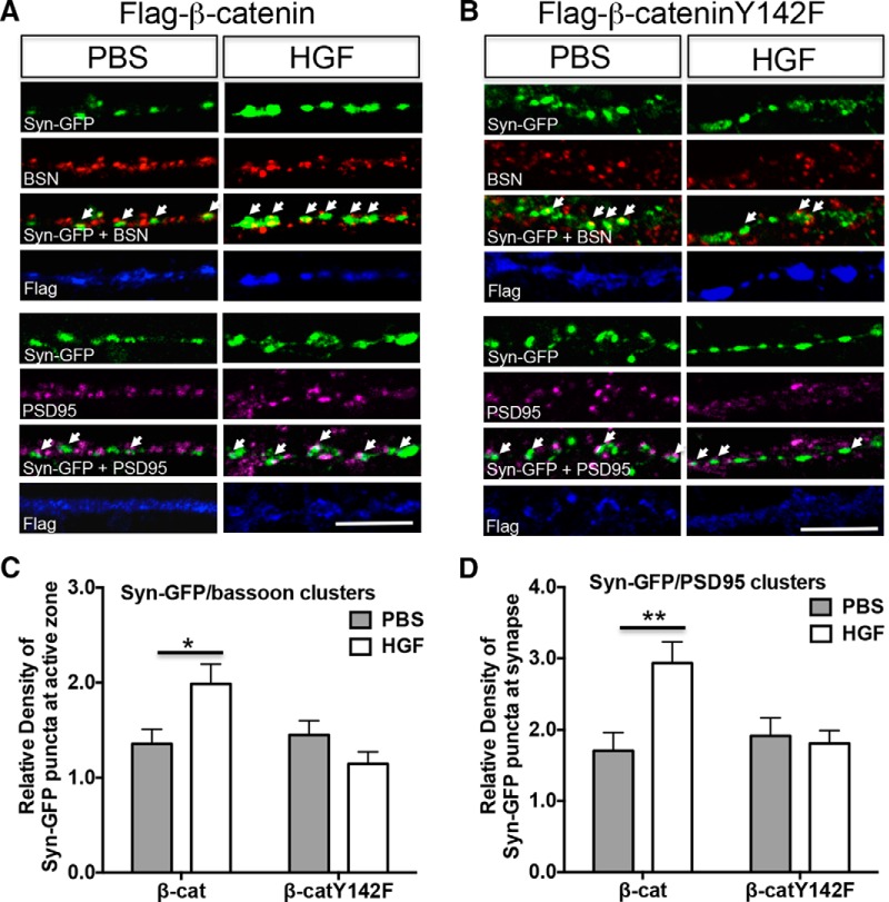
Figure 6.
Phosphorylation of β-catenin at Y142 is required for the HGF-induced increase in Syn-GFP/bassoon and Syn-GFP/PSD-95 clusters. A, B, Representative confocal microscopy images of Syn-GFP (green), bassoon (red), PSD-95 (magenta), and flag (blue) immunoreactivity in neocortical neurons at 14 DIV after treatment with PBS or HGF (25 ng/ml) for 10 min. The neurons were cotransfected with Syn-GFP and either flag-β-catenin (A) or flag-β-cateninY142F (B) plasmids at 5 DIV. White arrows indicate clusters colabeled with Syn-GFP/bassoon (top panels) or Syn-GFP/PSD-95 (bottom panels). Scale bars: A, B, 5 μm. C, D, Quantitative analysis of the density of Syn-GFP/bassoon (C) and Syn-GFP/PSD-95-colabeled clusters in β-catenin (β-cat)- or β-cateninY142F (β-catY142F)-transfected neurons. Each parameter was normalized in each culturing session to the mean value of Syn-GFP clusters in the PBS-treated group. Error bars represent the SEM; N = 24 cells from three independent culturing sessions for bassoon colabeling assay; N =30 cells from three independent culturing sessions for PSD-95 colabeling assay. Note that the inability to phosphorylate Y142 residue following HGF treatment results in no change in the colabeling of presynaptic/postsynaptic markers. *p < 0.05, **p < 0.01 (HGF vs PBS).

