Figure 4.
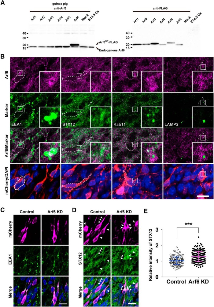
Arf6 localizes at endosomes and regulates the distribution of syntaxin12-positive endosomes in migrating neurons. A, Immunoblot analysis showing the specificity of rabbit anti-Arf6 antibody. The lysates of the mouse cerebral cortex (Cx) at E14.5 (10 μg) and HeLa cells transfected with the indicated FLAG-tagged Arf plasmids were immunoblotted with antibodies against Arf6 or FLAG. The positions and sizes of the molecular weight markers are indicated on the left. B, Representative micrographs showing endosomal localization of Arf6 in migrating neurons in the IZ at E17.5. Coronal sections of the E17.5 cerebral cortex, which had been electroporated with pCAGGS-mCherry at E14.5, were subjected to immunofluorescent staining with antibodies against Arf6 (magenta), the indicated markers (green), and mCherry (red). Nuclei were counterstained with DAPI (blue). Insets show the high-magnification views of boxed areas. C, D, Representative micrographs showing the subcellular localization of EEA1-positive (C) and syntaxin12 (STX12)-positive (D) endosomes in migrating neurons transfected with Control or Arf6 knockdown (Arf6 KD) plasmid in the IZ at E17.5. Arrowheads in D indicate STX12-positive puncta in transfected neurons. E, Quantification of the relative immunofluorescence intensity for STX12 in the perinuclear region. The relative immunofluorescence intensity was calculated by normalizing the immunofluorescence intensity for perinuclear STX12 in transfected neurons to that in the surrounding untransfected neurons. Note the significant increase in perinuclear STX12 in Arf6-KD neurons (Control, n = 155 cells; Arf6 KD, n = 239 cells). Data collected from three embryos in each condition were presented as mean ± SD and statistically analyzed with unpaired t test (*** P < 0.0001). Scale bars: B–D, 10 μm.

