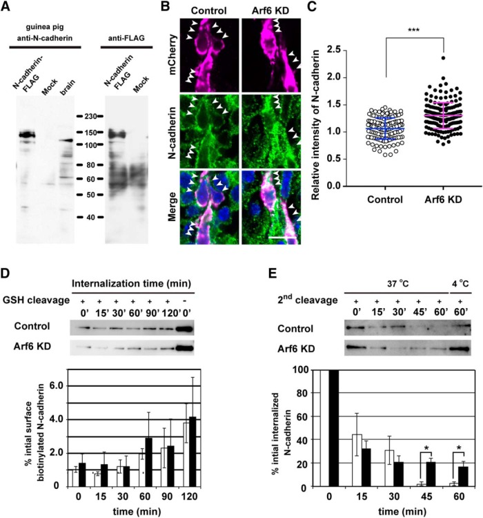
Figure 5.
Arf6 regulates endosomal trafficking of N-cadherin in cortical neurons. A, Immunoblot analysis showing the specificity of guinea pig anti-N-cadherin antibody. The lysates of the adult mouse brain (10 µg) and HeLa cells transfected with or without C-terminally FLAG-tagged N-cadherin were immunoblotted with antibodies against N-cadherin or FLAG. B, Representative micrographs showing the subcellular localization of N-cadherin in migrating neurons transfected with Control or Arf6 knockdown (Arf6 KD)in the IZ at E17.5. Arrowheads indicate the localization of N-cadherin in transfected neurons. C, Quantification of the relative immunofluorescence intensity for cytoplasmic N-cadherin. The relative intensity was calculated by normalizing the immunofluorescence intensity for cytoplasmic N-cadherin in transfected neurons to that in the surrounding untransfected neurons (Control, n = 137 cells from 3 embryos; Arf6 KD, 174 cells from 3 embryos). Note the significant increase in cytoplasmic N-cadherin in Arf6-KD neurons. D, Internalization assay for N-cadherin in cultured cortical neurons. A bottom graph shows the quantification of biotinylated N-cadherin internalized in neurons transfected with Control (open bars) or Arf6 KD(black bars; Control, n = 5 independent experiments; Arf6 KD, n = 5). E, Recycling assay for N-cadherin in cultured cortical neurons. A bottom graph shows the quantification of biotinylated N-cadherin retained inside neurons transfected with Control (open bars) or Arf6 KD (black bars; Control, n = 5 independent experiments; Arf6 KD, n = 6). Data were presented as mean ± SD in C and mean ± SEM in D and E, and statistically analyzed by unpaired t test in C (***P < 0.0001), and Mann-Whitney U test in D and E (*P < 0.05). GSH, Glutathione. Scale bar: B, 20 μm.

