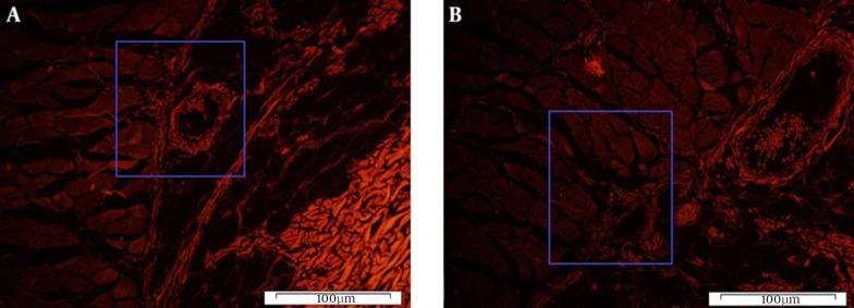
Figure 6. Histological Section of BM-MSC-Treated Skin Flap.
A, evidence of Dil fluorescence of BM-MSC-treated flap tissue in areas with high nuclear density (Dil) tissue on the transplant areas 7 days after flap elevation; B, the same section, but a different area. Note the close position of BM-MSCs to microvessels in the skin flap tissue (magnification ×100).

