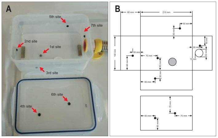
Fig. 1.
View of the interior of the container without its lid and a schematic diagram. (A) The photograph shows the interior of the food container without its lid. (B) The design schematic. The numbers are in order of biopsy progression and indicate the following: the greater curvature (GC)/anterior wall (AW) of the lower body (LB) (1st site, arrow), the AW of the antrum (2nd site, arrow), the AW of the LB (3rd site, arrow), the lesser curvature (LC)/posterior wall (PW) of the LB (4th site, arrow), the PW of the mid-body (MB) (5th site, arrow), the LC of the MB (6th site, arrow), and the cardia (7th site, arrow). The corresponding locations of the biopsy sites (●), polyvinylchloride (PVC) bottle cap (gray spot), and the hole for the scope shaft (○) are shown.

