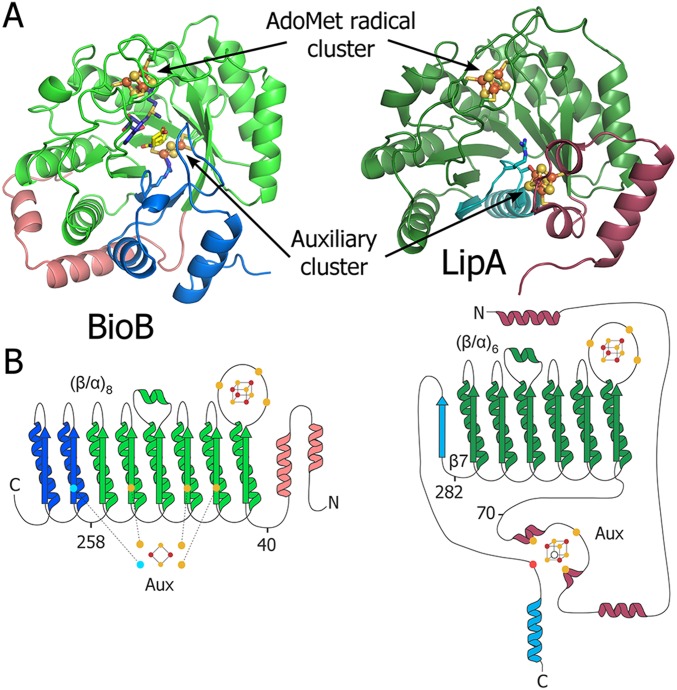
Fig. S1.
Overall fold of BioB and LipA. (A) Crystal structures of (Left) BioB and (Right) LipA in ribbon representation colored by domain. BioB adopts a (β/α)8 full triosephosphate isomerase (TIM) barrel fold, whereas LipA contains the (β/α)6 partial barrel more common among AdoMet radical enzymes, known as the AdoMet radical core fold. (B) Topology diagrams of (Left) BioB and (Right) LipA. Fe/S clusters are represented in ball and stick form. Fe/S cluster ligands are shown as circles and colored as follows: cyan, arginine; red, serine; and yellow, cysteine.

