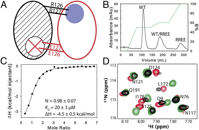
Fig. 1.
Characterization of the mixed dimers. (A) Schematic of a mixed labeled dimer showing the sites of mutation. The WT subunit (black) is 15N-labeled (hashed), and the RREE mutant subunit (red) is unlabeled. The blue circle and red X indicate the functional and nonfunctional binding site, respectively. (B) Chromatogram showing separation of the three species using anion exchange. (C) dUMP binding to the mixed dimer was measured by isothermal titration calorimetry (ITC); mean and SD are from three replicates. For the selected isotherm, mixed dimer concentration was 150 μM with 3 mM dUMP in the syringe. (D) Overlay of HSQC spectra of apo WT (black), WT-labeled (green), and RREE-labeled (red) mixed dimers. Residues proximal to the mutation are D124 and S125 in the RREE-labeled and L172 in the WT-labeled mixed dimers.

