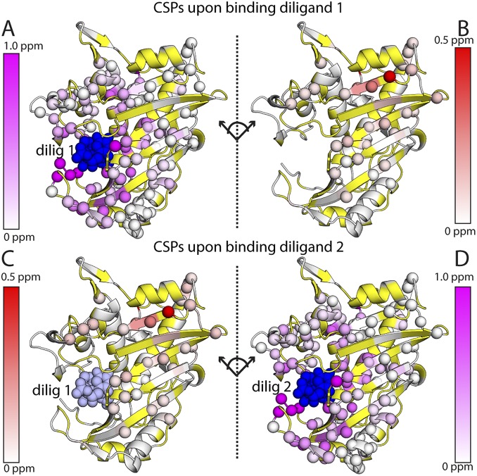
Fig. S6.
Chemical shift perturbations upon binding the first and second diligand. The effects of binding the first diligand to the binding (A) and nonbinding (B) subunits of the dimer are shown at the Top. The effects of binding the second diligand to the nonbinding (C) and binding (D) subunits are shown at the Bottom. Viewing the dimer interface region is enhanced by separating the two subunits, where the subunits on the right underwent a hinge-type rotation (dotted line) to yield the same viewing angle as those on the left. Residues with significant CSPs are shown as spheres. Residues that are missing or unassigned are in yellow. The first bound (dilig 1) and second bound (dilig 2) diligands are shown in dark blue; for binding of dilig 2 (Bottom), the previously bound dilig 1 is shown in light blue.

