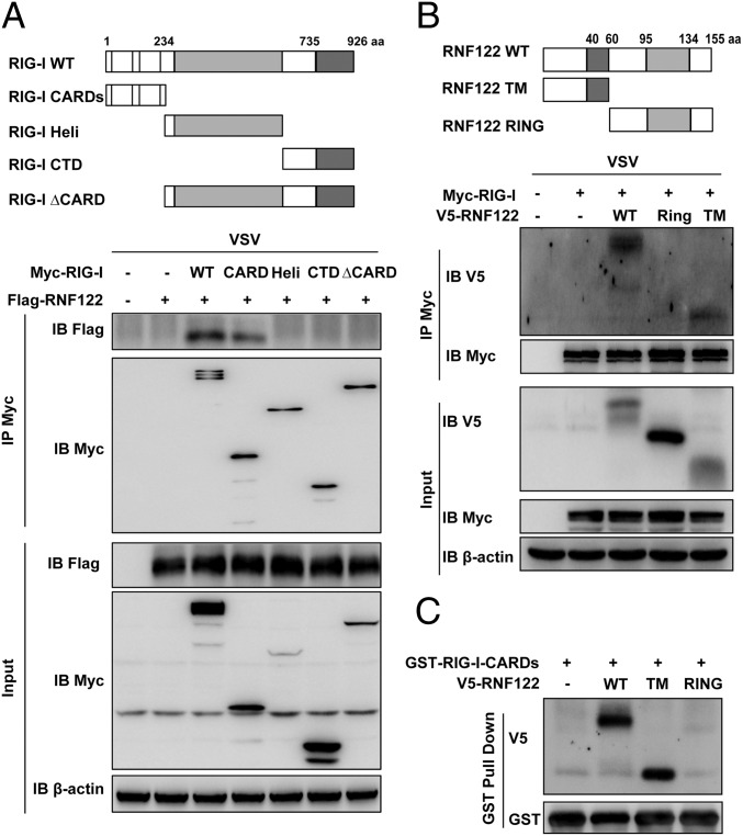
Fig. 5.
TM domain of RNF122 associates with the CARDs of RIG-I. (A and B) Schematic structure of RIG-I and RNF122 and the derivatives used in this work (Upper). HEK293T cells were transfected with the indicated plasmids and infected with VSV for 4 h. Cell lysates were immunoprecipitated by using anti-Myc antibody. The precipitates were analyzed by Western blot using indicated antibodies. (C) HEK293T cells were transfected with V5-RNF122 (WT), V5-RNF122-TM, or V5-RNF122-RING plasmids. After 24 h, cell lysates were incubated with purified GST-RIG-I CARDs proteins and subjected to GST pull-down assays. The proteins bound to glutathione–Sepharose beads were analyzed by Western blotting with the indicated antibodies.

